Research Featured on The Cover
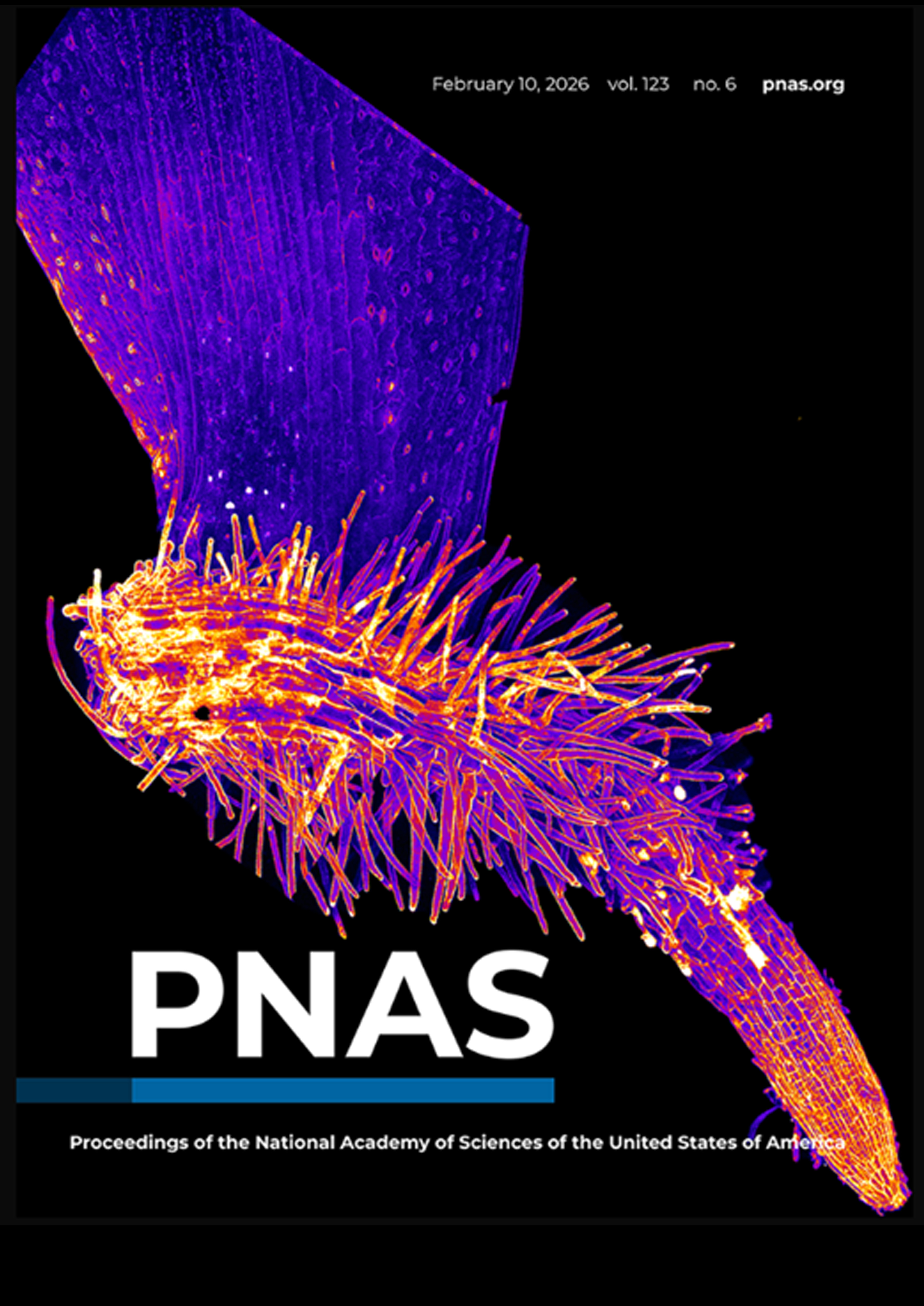
On the cover: Pictured is a regenerating root from an Arabidopsis thaliana leaf. Akansha Ganguly et al. detached plant leaves to study the mechanisms of stress mitigation associated with wound repair. The authors found that plant-specific PLETHORA transcription factors activate the ATG8 gene, which initiates recycling of damaged cell components to decrease intracellular stress and allow stem cells to differentiate. ATG8 is not activated if the plant is simply forming a callus, rather than regenerating roots. The results reveal how plant-specific autophagy regulators interact with intracellular stress signaling to aid in wound repair. See the article by Ganguly et al., e2513954123. Image credit: Akansha Ganguly.

On the cover: Progenitor progression into a complete plant system during de novo shoot regeneration is reimagined in a humanoid fashion. In this abstraction, a line connects the progressive stages of development from bottom-up. A glass shell (signifying the shell of cells expressing cell-wall loosening enzyme, XTH9 which generates a circumferential force field around the progenitor) encapsulates the young progenitor (juvenile plant in criss-cross position). This is until the progenitor has grown enough to break open the shell from within to emerge out and develop into a fully grown plant. The progression from slouched form with bud flower to upright form with flower in full bloom conveys the finer aspects of development.
Concept and drawn: Mabel Maria Mathew
Digitalized: Arun Kumar Kundu (Science Media, IISER-Pune)

On the cover: Artistic rearrangement of leaf confocal micrographs displaying battery of fluorescently tagged cell fate determinants in response to injuries. Images created by Anju P. S., Dhanya Radhakrishnan and Abdul Kareem V. K. Artwork by Mabel Maria Mathew. See Research article by Radhakrishnan et al. (dev185710).

On the cover: The cover image depicts the expression of multiple cell fate determinants and the PLT2 transcription factor gradient in growing root tips. In this issue, Durgaprasad et al. report that dosage of PLT2, expressed in a gradient, orchestrates regeneration competence at the root tip. Image by Kavya Durgaprasad and artwork by Anju P S

On the cover: Artistic representation of the journey of de novo shoot regeneration in Arabidopsis. Merged confocal micrographs show the expression pattern of YFP-tagged PLETHORA (PLT) transcription factors (yellow) in leaf explant (bottom) and in regenerating progenitors and shoot primordia after inductive cues (top). In this issue, Kareem et al. (pages 1017–1030) report that PLT-mediated regulatory modules uncouple the acquisition of competence to regenerate shoot progenitors from completion of shoot regeneration, indicating a two-step mechanism for de novo shoot regeneration. Image by Abdul Kareem.

On the cover: Regenerating shoot from lateral root primordia in Arabidopsis. Merged confocal micrographs show the expression pattern of GFP tagged polar auxin efflux carrier PIN-FORMED1 (PIN1) membrane protein (green) and venus-YFP tagged auxin response sensor DR5 (yellow) in shoot primordia after inductive cues. In this issue, Kareem et al. (Regeneration 2016; 3(4):182–197) review the recent advances in understanding the molecular mechanism of de novo plant regeneration. Image by Abdul Kareem at IISER

On the cover: Santuari et al. reveal how the PLT gradient regulates cell state by region-specific induction of cell proliferation genes and repression of differentiation. The cover image shows how PLT2 expression influences the boundary of meristem target gene expression domains at the root tip. Activation of ProHAN:3xvYFP expression in a transgenic root after 0, 6, and 24 h of DEX induction of PLT2-GR expressed under control of the constitutive 35S promoter. Ectopic induction is obvious at 6 and 24 h of transfer to DEX media from the enhanced expression of fluorescent protein throughout the root outside of the meristematic domain of endogenous PLT2 expression. This image series shows that PLT levels determine the proximal/shootward boundary of the target gene expression domain, consistent with a dose-dependent role in zonation.
Research Highlights/Blog


Plants Rebuild Roots Using Geometry, Not Just Genes: Study
Scisoup, September 03, 2025

How plant roots rebuild their tips after injury
Nature, September 09, 2025

Research in News
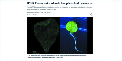


Biologists of IISER-Pune decode 'cellular secret talk'...
The Indian Express, September 8, 2022

How wounded plants heal survive
The Hindu, March 07, 2020

Decoding how plant roots regenerate
The Hindu, October 19, 2019

Top journal hails research on plant regeneration
Deccan Chronicle, November 18, 2015

City Scientists Unravel Mystery of Plant Regeneration
The New Indian Express, November 17, 2015

Study led by IISER Pune decodes mechanisms…
The Indian Express, July 28, 2025

Reshaping during healing in plants
The Hindu, August 23, 2025

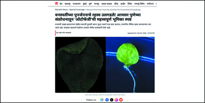
Saamna, August 10 2025